


Neuronal Protein Tracing Reveals How the Brain Routes Its Waste

The brain is one of the busiest organs in the body, constantly processing and reshaping itself. That activity produces an equally constant stream of molecular byproducts—proteins that need to be moved out before they accumulate. When those clearance routes slow or break down, waste lingers, and the consequences can be profound. In Alzheimer’s disease, for example, toxic proteins build up in vulnerable regions. Yet despite decades of research, scientists have lacked a clear view of how waste normally leaves the brain.
A new study from the Gladstone Institutes offers the clearest picture yet of how the brain normally takes out its trash—and what happens when those routes fail. Published in Cell as “Physiological brain clearance architecture revealed by neuronal protein tracing,” the work introduces a method that traces waste proteins from the moment they are produced inside neurons to the moment they leave the brain.
For decades, researchers have relied on injecting tracers into the cerebrospinal fluid (CSF) to visualize drainage. But this approach, while illuminating, shows all possible routes, instead of pinpointing the most-used exit. “These injected tracers disturb the very system we’re attempting to measure,” said lead author Andrew Yang, PhD, a Gladstone investigator. “We wanted to find a better way.”
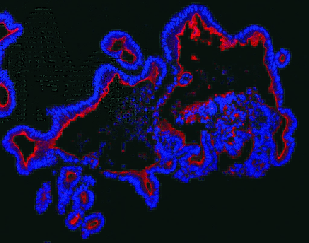
Yang’s team engineered neurons in mice to produce a fluorescent protein, ZsGreen, that could be followed as it exited the brain through its natural routes. This allowed the researchers to track waste as it moved into the dura, skull, nasal cavity, and lymph nodes—regions populated by specialized immune cells that interact with brain‑derived proteins.
The resulting map diverged sharply from the field’s long‑held assumptions. Traditional CSF tracers had pointed to the cervical lymph nodes as a major drainage site. But the new method revealed that very little neuronal waste actually reaches those nodes. “We were surprised to find that very little ZsGreen drained to the cervical lymph nodes,” Yang said. “Instead, waste drained through the dura, skull, and nasal cavity. Our findings underscore why tracking waste proteins themselves, rather than movement of the cerebrospinal fluid, provides a more accurate understanding of waste clearance dynamics.”
The team also uncovered a striking organizational principle: where a protein is made determines where it drains. Proteins produced in upper forebrain regions exited through upper routes, while those from deeper structures, such as the striatum, used lower pathways. The researchers call this the brain’s “nearest‑exit” model. “It’s like each brain region has a biological ZIP code system to ensure waste will be sent to the correct drainage site,” said Nalini Rao, PhD, a postdoctoral fellow. She noted that in aging or disease, these ZIP codes may become scrambled, potentially explaining why certain regions are more vulnerable to disorders like Alzheimer’s.
Disease models reinforced the system’s fragility. In mice with acute inflammation, ZsGreen leaked directly into the bloodstream, bypassing normal routes. In an Alzheimer’s model, waste became trapped inside the brain, unable to drain effectively. “Understanding how diseases disrupt brain clearance could help us design therapeutics to target the brain border compartments and enhance waste removal,” Rao said.
With their new tracing method, Yang’s group plans to probe how clearance changes across aging, sleep, and disease—and whether brain tumors exploit these pathways to evade immune detection. The architecture of brain waste disposal, once opaque, is now open for exploration.
The post Neuronal Protein Tracing Reveals How the Brain Routes Its Waste appeared first on GEN - Genetic Engineering and Biotechnology News.
Apa Reaksi Anda?
 Suka
0
Suka
0
 Kurang Suka
0
Kurang Suka
0
 Setuju
0
Setuju
0
 Tidak Setuju
0
Tidak Setuju
0
 Bagus
0
Bagus
0
 Berguna
0
Berguna
0
 Hebat
0
Hebat
0