


First 3D Structure of Malaria’s “Moving Junction” Solves Infection Mystery

For nearly half a century, scientists have known that malaria parasites force their way into human red blood cells (RBCs) through a ring-shaped structure called the moving junction (MJ). What no one could work out was what it actually does. The structure assembles, does its job, and dissipates in the space of 60 seconds—gone before anyone can get a close look.
A team at Columbia University has now finally caught the moving junction in the act. By freezing parasites at the onset of invasion and lifting the intact complex straight out of the cell, the researchers obtained the first high-resolution view of its three-dimensional structure. What they saw overturned a decades-old assumption about how the parasite gets in. Rather than a passive doorway, the moving junction turns out to be a molecular machine that actively remodels the host cell’s membrane to help the parasite force its way inside.
The findings detail how the team obtained the structure and then used it as a blueprint to design a mini-protein, from scratch, that blocks invasion—a proof of concept for a new kind of antimalarial drug.
“We’ve known for decades that this structure is essential for the parasite to get into a cell, but not how it actually works,” said Chi-Min Ho, PhD, an assistant professor in the Department of Microbiology and Immunology at Columbia University Vagelos College of Physicians and Surgeons and the study’s senior author. “Pulling it directly out of the parasite intact let us finally ask that question directly.”
Ho is senior author of the team’s published paper in Cell, titled “Structural basis for host membrane binding and remodeling by invading malaria parasites.” In their paper, the team stated in summary, “This work represents a major step toward resolving the decades-long mystery surrounding the structure and function of the malarial MJ, underscoring the power of pursuing native structures and laying the foundation for structure-guided design of next-generation antimalarials.”
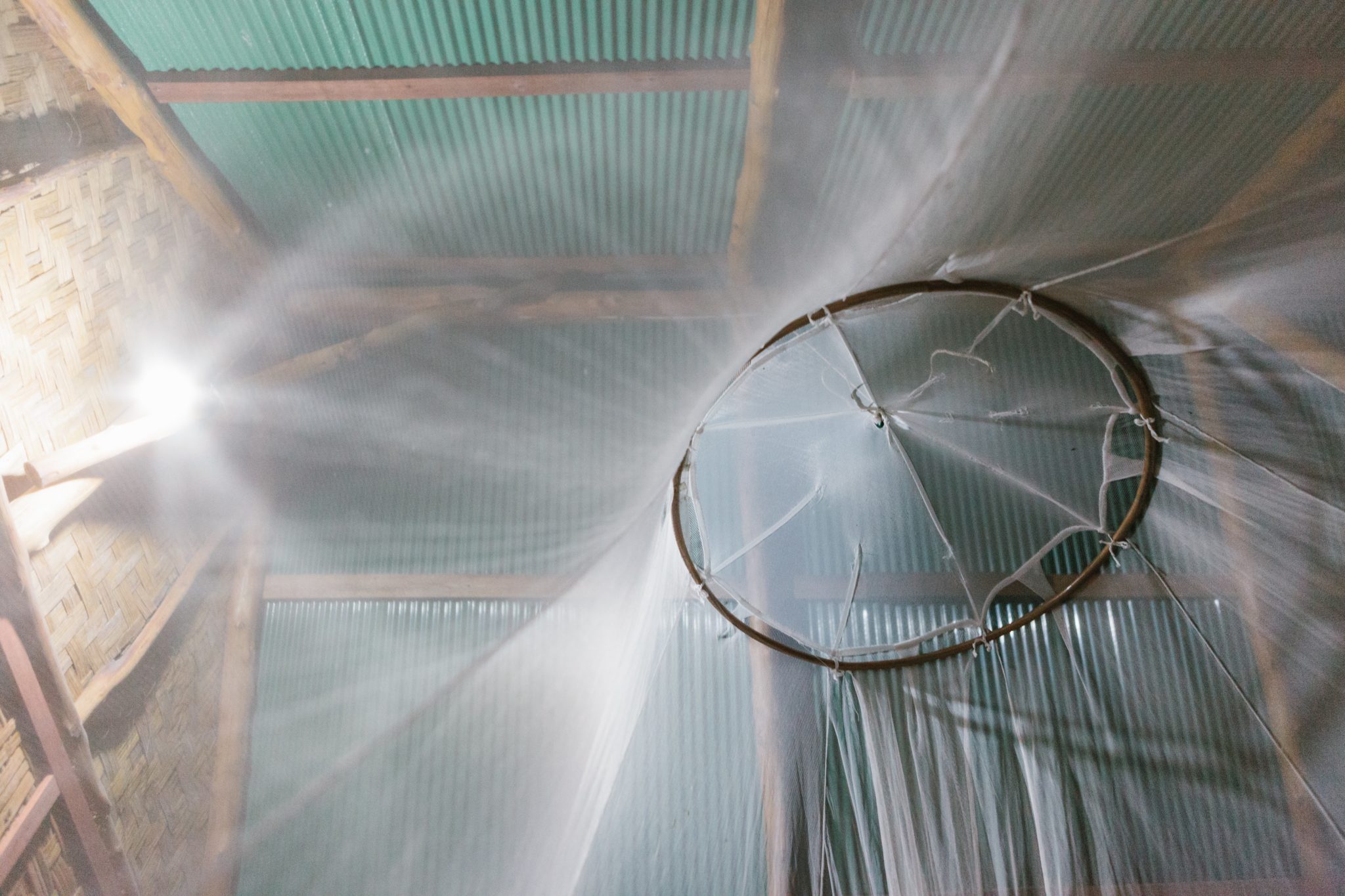
Malaria still kills roughly 600,000 people a year, the overwhelming majority of them young children in sub-Saharan Africa, and the parasite is steadily becoming resistant to frontline drugs. “Malaria morbidity and mortality are directly linked to the invasion and replication of the malaria parasite Plasmodium falciparum in human red blood cells (RBCs),” the authors wrote. The malaria parasite life cycle involves two hosts, humans and Anopheles mosquitoes, and infecting human RBCs and hepatocytes, as well as mosquito salivary glands.
The disease starts with a single event: a parasite breaking into a red blood cell. “Parasites establish infection by invading host cells in a rapid and precisely choreographed process …” the team continued. In an infected person, trillions of parasites are released and invade every 48 hours in synchronized waves. This rhythmic cycle of rupture and reinvasion drives the periodic fevers malaria is known for. “After gliding, reorientation, and initial attachment, parasite internalization is initiated by the formation of a ring-shaped ultrastructure called the moving junction (MJ), which anchors the parasite to the host cell,” the researchers explained.
The same moving junction machinery is used across every species and every stage of the parasite’s life cycle, which has made it one of the most sought-after targets in malaria research. For antimalarial drug and vaccine development, block it, and you stop infection at its source.
The moving junction has been a puzzle since 1978, when scientists first observed in electron microscopy images a mysterious thickening of the membrane where parasite meets cell. Researchers eventually identified the four parasite proteins—AMA1, RON2, RON4, and RON5—that assemble into the junction’s basic building block, and confirmed that all were essential for invasion. But what the structure actually did remained unknown, because it survives for a minute or so and refuses to reassemble in a test tube. “Efforts to address this critical gap in understanding have been thwarted by the short-lived (60–90s) nature of the complex, as well as by the difficulty of recapitulating it in heterologous systems for detailed biochemical and structural study,” the researchers stated.
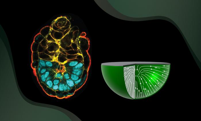
The Columbia team got around this by stopping invasion mid-stride. Using a compound that halts the parasite’s internal motor without preventing the junction from forming, they stalled parasites partway into red blood cells, then extracted the fully assembled AMA1-RON complex—the building block from which the whole junction is constructed—and imaged it with cryo-electron microscopy (cryo-EM), a technique where molecules are flash-frozen and imaged with an electron beam at extremely high magnifications to reveal their shape in atomic detail. The result was a sharp, three-dimensional view of that building block. The researchers noted that it was quite strikingly shaped like a sailboat, with the AMA1 protein forming a “sail” above the cell surface and the three RON proteins forming a broad “hull” pressed against the membrane below.
The biggest surprise was in the hull, where the team found clues that finally hinted at the moving junction’s role in invasion. The face of the structure pressed against the host membrane is blanketed with positively charged anchors, and the surface is studded with short helices that drive deep into the membrane like wedges. “These short helices insert asymmetrically into one leaflet of the membrane, displacing lipid headgroups and applying lateral pressure to generate local membrane deformations.”
Both features are widely recognized hallmarks of a well-known family of cellular machines that bend and reshape membranes. Their structural findings, they noted in their report, reveal “a highly unusual molecular staple that exhibits the hallmarks of a powerful membrane-remodelling machine.”
To test whether the structure could indeed deform a membrane, the researchers synthesized the parasite’s wedge-like helices and added them to artificial membrane bubbles. The membranes thinned and punctured. Meanwhile, weakened versions of the helices left the bubbles intact. The team concluded that the moving junction appears to pull the host membrane into shape, likely working in concert with the parasite’s motor to lever the parasite inside.
“It had been pictured as a kind of series of staples or spot-welds, making up a passive ring the parasite hauls itself through,” said Meseret Haile, the study’s first author and a PhD candidate in Ho’s lab. “What we see instead is a machine built to reshape the host cell’s own membrane. That changes how we think about the whole event.” In their paper, the team added, “Our work reveals that, although visually suggestive of canonical tight junctions, the MJ differs fundamentally in function, serving as a dynamic portal that orchestrates parasite internalization, rather than a static adhesion molecule.”
Beyond finally revealing how the moving junction allows the parasite to invade, the structure also gave the team a precise map of where and how AMA1 grips its partner protein, the contact that holds the entire junction together. Using a machine learning-powered protein-design tool together with their structural information, the researchers designed a mini-protein to break that grip. Their best candidate blocked parasites from invading red blood cells in a dose-dependent way and left already-infected cells unaffected, confirming that it works specifically by stopping entry rather than through general toxicity.
The designed mini-protein is a first proof of concept, not a drug, and will need considerable refinement before it could be tested in people. But it demonstrates an exciting new strategy: using near-native structures to design invasion-blocking mini-proteins against a target that has long frustrated conventional approaches. The same structure also clarifies how several leading anti-malaria antibodies work, information that could feed back into vaccine design. “Our successful proof of principle demonstrates the potential power of context-driven binder design for challenging systems, offering a previously unexplored avenue for therapeutic intervention,” they wrote. “In addition to their therapeutic potential, these binders may also serve as powerful tools for probing the functional relevance of specific protein interactions.”
Daphne Kaxiras, an MD-PhD student in Ho’s lab who led the inhibitor design, said, “Once we could see the target in its real setting, designing something to block it became a tractable problem. That’s the part we’re most eager to build on.”
The team’s approach, imaging fragile complexes captured directly from the organism and using them to guide design, may apply to many other parasites and pathogens that are notoriously difficult to study.
The post First 3D Structure of Malaria’s “Moving Junction” Solves Infection Mystery appeared first on GEN - Genetic Engineering and Biotechnology News.
Apa Reaksi Anda?
 Suka
0
Suka
0
 Kurang Suka
0
Kurang Suka
0
 Setuju
0
Setuju
0
 Tidak Setuju
0
Tidak Setuju
0
 Bagus
0
Bagus
0
 Berguna
0
Berguna
0
 Hebat
0
Hebat
0