

















































%20and%20Batch%20Production%20Records%20(BPR).png)















Spatial Biology Moves Toward Translation
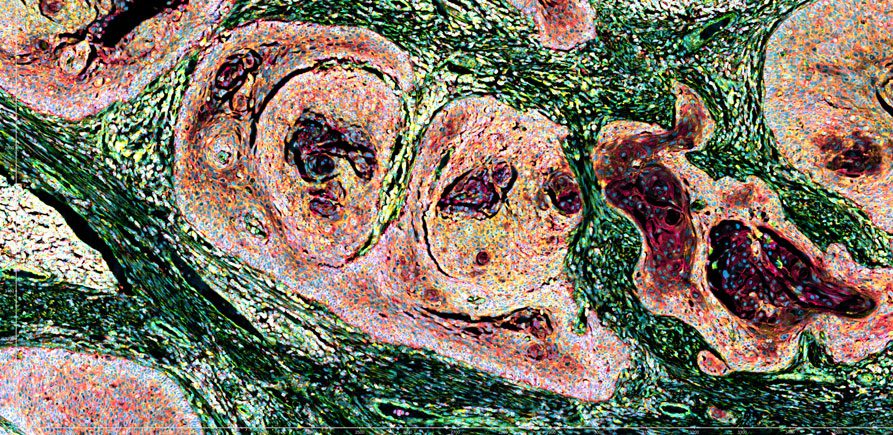
If “Spatial Biology” conjures images of a science conducted in zero gravity aboard the International Space Station, think again. As one of the most rapidly advancing areas in biomedical research and precision medicine, spatial biology probes the inner landscape of intact cells and tissues down to single-molecule resolution. By preserving location and organization, instead of dissociating tissue into component parts, this discipline maps fine architecture, cellular neighborhoods, and individual molecules.
Maintaining that context is driving new ways of characterizing disease progression, identifying biomarkers, advancing therapeutics, and monitoring treatment responses. Fueling these advances is an expanding toolbox that encompasses high-plex imaging, spatial transcriptomics and proteomics, multiomic profiling, and increasingly sophisticated computational analyses. We consulted industry leaders about how the field is progressing, barriers that remain, and their vision for what is needed to propel spatial biology into its next phase.
Scaling spatial discovery
While genomics and single-cell sequencing profile molecular features and cellular states, spatial biology answers questions that neither can address, such as where those features and cells are located and how they are organized.

Senior Director
10x Genomics
“When you dissociate a tissue into individual cells, you lose something fundamental: where those cells were and how they were organized relative to one another,” observes Kamila Belhocine, PhD, senior director of spatial biology at 10x Genomics. She continues: “Spatial biology puts that context back. In cancer, for example, knowing that a particular immune cell population is present within a tumor sample is useful. But knowing whether those cells have infiltrated the tumor core, are sitting at its margin, or are excluded from it altogether can tell you something very different about the biology, and potentially, about response to treatment. The same principle applies to understanding tissue architecture in cases like development and cell differentiation, and disease progression.”
Several barriers have prevented spatial biology from going mainstream in the biopharma/biotech industry. Belhocine says, “I think about four things: speed, reliability, cost, and interpretability. Spatial needs to be fast enough to fit into routine research workflows, robust enough to generate consistent results at scale, affordable enough to use across large studies and, ultimately, straightforward enough to turn the resulting data into biological insight. The first three are fundamentally technology and engineering challenges. Interpretability is harder because it also requires accumulating evidence and validating which spatial signatures are truly meaningful and actionable.”
However, according to Belhocine, single-cell technologies had to address similar issues. “A decade ago, single-cell analysis was largely a specialized technique. Continued innovation made it easier, more scalable, more robust, and dramatically more cost-effective, opening it to a much broader community and enabling much larger studies. We think spatial is following a similar trajectory.”

Chief Technology Officer
Bio-Techne
Belhocine says the company’s newest spatial instrument, Atera, which will be shipping in the second half of the year, is an important step in that progression. “This technology combines whole-transcriptome spatial profiling with single-cell sensitivity and subcellular resolution at a massive scale designed for much larger studies.”
Looking ahead, Belhocine predicts, “As spatial technologies become more scalable, robust and cost-effective, researchers can apply them across much larger patient cohorts to identify biomarkers and signatures of treatment response. Additionally, AI will undoubtedly accelerate that trajectory even further.”
Spatial multiomics
Gary Latham, PhD, CTO, Bio-Techne, provides another perspective on the role of integrating protein analysis and gene expression in spatial biology. “A fuller understanding of tissue biology requires seeing where proteins and genes are expressed, how they relate, and how they interact within the tissue architecture.”
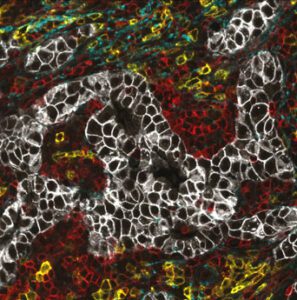
This molecular multiplexing can provide a more complete view of tissue biology. “Spatial biology is unique from other technologies because it takes us from seeing what is there to understanding what is where,” says Latham. He continues, “This is critical because biology can’t be understood as a parts list. Instead, it is a dynamic system of interactions and networks where location defines meaning. While single-cell sequencing can tell you which cell types exist and their molecular states, spatial biology tells you how those cells are organized, how they may be interacting, and how these relationships may influence disease biology and clinical outcomes.”
Latham indicates that the field is working to overcome several challenges. “There is a need for reproducible, standardized automation for today’s biomarkers and tomorrow’s multiomic precision medicine. Many spatial biology workflows remain exploratory and manual during early discovery phases. As programs move toward translational research, the focus shifts to robustness, reproducibility, and standardized, validated workflows. Ultimately, clinical adoption requires validated panels, automation, and throughput suited to routine testing.”
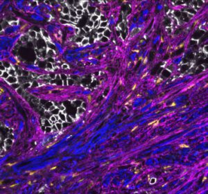
According to Latham, Bio-Techne is helping address these barriers in several ways. Its COMET Suite provides an end-to-end automated platform for hyperplex immunofluorescence and multiomics staining, imaging, and analysis of tissue samples. Additionally, its RNAscope portfolio enables same-section multiomics, with 80,000 RNA probes spanning 450 species and compatibility with multiple automated staining instruments.
Latham believes the spatial multiomics approach can connect gene and protein expression with tissue architecture within the same cells and microenvironment, revealing biological relationships and linking state to function that no single modality can uncover.
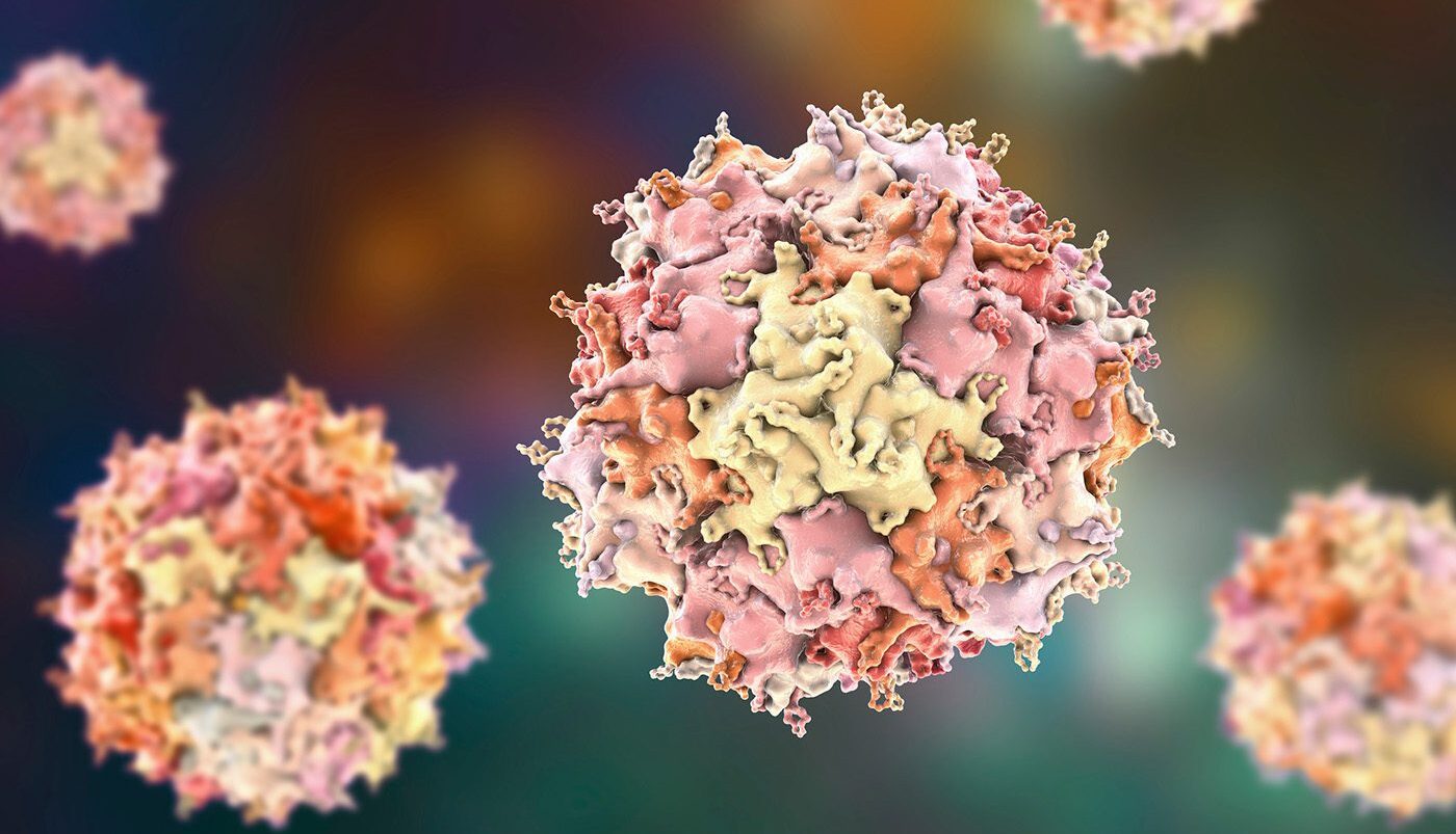
He says several areas are seeing the fastest adoption of spatial biology. “Oncology and immuno-oncology continue to dominate spatial biology applications, while immunology and neuroscience represent the next-most prominent areas of focus. Biodistribution of gene therapies is an especially high-impact application: spatial multiomics can localize an AAV vector, transgene mRNA, or oligonucleotide therapeutics such as ASOs and siRNAs to specific cells, then measure changes in the target or downstream gene expression and the associated protein phenotype.”
Integrated discovery & translation
Spatial biology is increasingly moving beyond simply identifying where cells are located to understanding how cellular neighborhoods, cell–cell interactions, and biological pathways shape disease biology. As Thomas Campbell, PhD, senior director of product management for Bruker Spatial Biology, explains, “Biology is inherently multi-dimensional, and no single spatial technology can or will capture every aspect of that complexity. The field needs multiple fit-for-purpose approaches that allow researchers to ask different questions and connect insights across DNA, RNA, proteins, and cellular function.” This ability to examine biology from complementary perspectives is becoming increasingly important as spatial research moves from discovery toward translation.
According to Campbell, understanding these context-dependent differences is driving the field. “The next phase of spatial biology will be about using high-plex discovery to develop focused, quantitative assays that can be deployed across large cohorts. One of the biggest challenges in translational biopharma is the discovery-to-translation gap. A study may identify dozens or hundreds of potentially relevant biomarkers, but a translational or clinically deployable assay demands a much smaller, highly reproducible panel.”
Thus, defining a clinically relevant spatial signature requires insights across the genome, transcriptome, and proteome. Campbell says Bruker Spatial Biology’s portfolio comprises platforms spanning multiple omic layers—including DNA, RNA, proteins, post-translational modifications, miRNA and live cell function—“because we recognize that no single modality can fully resolve the biological complexity that drives disease.”

Senior Director
Bruker Spatial
That integrated approach is reflected in several of the company’s platforms. CosMx® Spatial Molecular Imager (SMI) provides a view of the whole transcriptome at subcellular resolution, while enabling the simultaneous measurement of RNA and protein within the same tissue section. The CellScape XR platform provides quantitative spatial proteomics using flexible assay chemistry. Meanwhile, the GeoMx® Digital Spatial Profiler (DSP) profiles the whole transcriptome and more than 1,200 proteins from distinct tissue compartments. Campbell says that, together, these three platforms “provide complementary views of biology and, when used together, enable a more complete understanding of cellular function across multiple omic layers.”
Campbell sees this transition from discovery to translation as one of the most important opportunities for spatial biology. “Whether it’s in oncology, immunology, or neuroscience applications, the future of spatial biology is not simply just about generating more data; it’s translating increasingly sophisticated spatial discoveries into assays that can answer focused biological and clinical questions across large numbers of samples,” he says. “That requires a portfolio of complementary technologies. High-plex discovery platforms can reveal the complexity and heterogeneity of disease, while more focused and quantitative approaches can translate those findings into reproducible biomarkers and clinically relevant assays. Ultimately, the value of spatial biology will be measured by how effectively we can connect discovery to clinically actionable insights.”
From plex to precision
“Historically, spatial biology involved a fundamental tradeoff between plex, sensitivity, and resolution—researchers had to sacrifice one to gain another,” suggests Mike Miller, PhD, CTO and CPO at Quanterix. He says that is now shifting. “With today’s tools, those tradeoffs have narrowed considerably. The current challenge becomes building a platform that delivers reproducibility, rigor, and clinical-grade integrity at scale.”
The company is addressing those needs in several ways. Miller reports: “Our Akoya PhenoCycler-Fusion platform enables researchers to visualize and quantify 100+ proteins in a single tissue section at single-cell resolution.” The platform uses molecular barcoding chemistry. Miller elaborates: “Molecular barcoding is a way of identifying and measuring multiple proteins in a tissue while avoiding many of the limitations of traditional immunohistochemistry and conventional multiplexing approaches.”

Chief Technology Officer
Quanterix
This approach combines three main components: an antibody that recognizes the target of interest; a unique molecular barcode, consisting of a short oligonucleotide sequence attached to the antibody; and a complementary oligonucleotide carrying a fluorescent reporter that binds to the barcode, enabling target visualization and quantification. “The key advantage is that the fluorescent reporter can be reversibly hybridized and removed without removing the antibody or repeatedly staining and stripping the tissue. This enables a cyclical imaging workflow.”
The company also recently introduced its PhenoCode2.0 chemistry as part of its new molecular barcoding kit, “which lets researchers reliably barcode virtually any antibody of their choice.” Miller continues: “This means researchers can easily expand and customize their biomarker panels to address new hypotheses, incorporate emerging biomarkers, and scale between unbiased discovery and targeted validation, accelerating the path to new insights.”
Beyond expanding what can be measured, Miller believes the next phase of spatial biology will focus on translating high-plex discoveries into larger, clinically relevant studies. “That’s ultimately what will separate platforms going forward, bridging the gap between generating a striking image in discovery and generating a measurement that holds up in a clinical trial or diagnostic setting.”
The post Spatial Biology Moves Toward Translation appeared first on GEN - Genetic Engineering and Biotechnology News.
Apa Reaksi Anda?
 Suka
0
Suka
0
 Kurang Suka
0
Kurang Suka
0
 Setuju
0
Setuju
0
 Tidak Setuju
0
Tidak Setuju
0
 Bagus
0
Bagus
0
 Berguna
0
Berguna
0
 Hebat
0
Hebat
0