


Single-Cell Atlas of the Prenatal Brain Reveals How Down Syndrome Reshapes Development
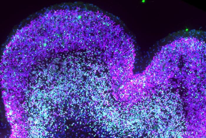
A cellular-resolution molecular map details how Down syndrome alters human brain development before birth. The study analyzed more than 100,000 nuclei from human prenatal neocortex samples collected across 26 pre-genotyped donors during gestational weeks 13 to 23—the only window during which all the cortical neurons a person will carry for their entire life are generated. The findings suggest that Down syndrome disrupts the developmental sequence of that process, creating shifts that may help explain later differences in cognition, learning, and sensory processing.
This work is published in Science in the paper, “A single-cell multiomic analysis identifies molecular and gene-regulatory mechanisms dysregulated in developing Down syndrome neocortex.“
“There’s a new level of detail here that had never existed before,” said Luis de la Torre-Ubieta, PhD, an assistant professor of psychiatry and biobehavioral sciences at UCLA and a member of the Eli and Edythe Broad Center of Regenerative Medicine and Stem Cell Research. “For the first time, we can really try to understand systematically what’s going on in the developing brain of individuals with Down syndrome.”
“No one had looked at the developing human brain in Down syndrome directly using single-cell genomics,” he continued.
The Down syndrome research field has historically focused on two areas: the adult brain and the disorder’s connection to neurodegeneration. What remained largely unexamined, despite clear indicators that Down syndrome is a developmental condition, was how the condition shapes the developing brain itself.
The development of the prenatal neocortex typically follows a tightly orchestrated sequence. Progenitor cells must first divide repeatedly to expand their own pool, building up a sufficient base for all future neurons. Only then do they begin differentiating into neurons, starting with deep-layer cell types and progressing toward upper-layer cells in a carefully timed order.
The study found that progenitor cells appear to rush prematurely into neuron production, depleting their own pool and skewing the balance of neuron types generated. Specifically, the researchers observed a relative increase in upper-layer intratelencephalic neurons and a reduction in deep-layer corticothalamic neurons.
Those two cell populations play fundamentally different roles: CT neurons project outward from the cortex—connecting to brain structures and to the spinal cord to govern sensation and movement; IT neurons wire within the cortex, connecting the two hemispheres and contributing to information processing. This finding offers a new hypothesis for how early developmental changes might contribute to the cognitive profile of the condition.
The finding also offers a new answer to a longstanding question in the field: Why do people with Down syndrome tend to have smaller brains? Earlier theories centered on elevated rates of cell death. The current study found less evidence of widespread neuronal death and instead points to the depletion of the progenitor pool.
The study employed paired single-nucleus multiomics to reconstruct not just a snapshot of which cells are present, but the regulatory programs that guide cell fate—and how those programs are disrupted in Down syndrome. Systems-level approaches also led them to uncover alterations in cell metabolism and changes in how the vasculature interacts with the developing nervous system, both of which could speed up neuron production.
The study’s significance extends beyond Down syndrome. The researchers specifically tested for overlap between the molecular disruptions they identified and the genetic risk signatures associated with other neurodevelopmental and neuropsychiatric conditions, including autism, epilepsy, and developmental delay. They found substantial convergence, particularly in the gene-regulatory networks governing the specification of IT versus CT neurons.
“Down syndrome could be a model to understand intellectual disability and neuropsychiatric disorders more broadly,” de la Torre-Ubieta said. “Also to uncover the shared biology underlying these conditions—because the mechanisms are often still unknown.”
The publication coincides with a companion paper from researchers at the University of Wisconsin-Madison, appearing in the same issue of Science. While the UCLA study focuses on the prenatal period, the Wisconsin team examined the postnatal brain, studying Down syndrome between approximately one and five years of age.
Together, the two papers provide a continuous molecular view of Down syndrome brain development from mid-gestation through infancy—a resource that did not previously exist and that the researchers expect will serve as a reference for their field for years to come.
While the researchers are careful to emphasize that the findings do not point to a near-term clinical application, the study provides the clearest picture yet of the cellular and molecular events that distinguish the Down syndrome brain during development, and a framework for identifying future therapeutic targets.
The post Single-Cell Atlas of the Prenatal Brain Reveals How Down Syndrome Reshapes Development appeared first on GEN - Genetic Engineering and Biotechnology News.
Apa Reaksi Anda?
 Suka
0
Suka
0
 Kurang Suka
0
Kurang Suka
0
 Setuju
0
Setuju
0
 Tidak Setuju
0
Tidak Setuju
0
 Bagus
0
Bagus
0
 Berguna
0
Berguna
0
 Hebat
0
Hebat
0