





























Double trouble: a case study on concurrent tetratrichomoniasis and coccidiosis in a com-mercial turkey operation
Gut health is a critical determinant of successful turkey production, directly influencing growth performance, feed conversion efficiency, and overall flock health. Disruptions to intestinal integrity frequently manifest as enteric disease complexes, leading to significant morbidity and economic attrition. Although various pathogens are known to affect the gastrointestinal tract of turkeys, maintaining optimal gut health under field conditions remains challenging due to the multifactorial nature of enteric disorders. This report describes an enteric disease outbreak in a commercial turkey flock where diagnostic investigation identified a co-infection of Tetratrichomonas gallinarum and a diverse profile of Eimeria species.
Introduction
Enteric diseases in turkeys are frequently multifactorial and may be associated with viral, bacterial, parasitic, or unidentified etiologies (1,2,3). Key viral contributors include Turkey Coronavirus (TCoV), the causative agent of Bluecomb disease; Hemorrhagic Enteritis Virus (HEV); and the multi-agent Turkey Poultry Enteritis Mortality Syndrome (PEMS), frequently associated with astroviruses. These viruses compromise the mucosal barrier, providing a portal for secondary bacterial pathogens.
Bacterial enteric diseases are also important contributors to intestinal pathology in turkeys. Necrotic enteritis, primarily caused by Clostridium perfringens, is a significant bacterial condition that can lead to severe intestinal damage, mucosal necrosis, and elevated mortality in affected flocks.
Parasitic burdens further complicate the enteric landscape. Heavy infestations with roundworms such as Ascaridia dissimilis can cause intestinal irritation, impaired nutrient absorption, and decreased growth performance. However, protozoal infections remain the most prevalent parasitic challenge in commercial production. Protozoal pathogens include Eimeria species causing coccidiosis, Histomonas meleagridis, the causative agent for histomoniasis, and Cochlosoma anatis, the causative agent of cochlosomiasis. These pathogens can occur individually or in combination, often resulting in complex enteric disease syndromes.
Given the complexity of enteric diseases in turkeys, accurate diagnosis through microscopic and molecular methods is essential for identifying the causative agents and implementing appropriate control strategies.
Case report
Case history
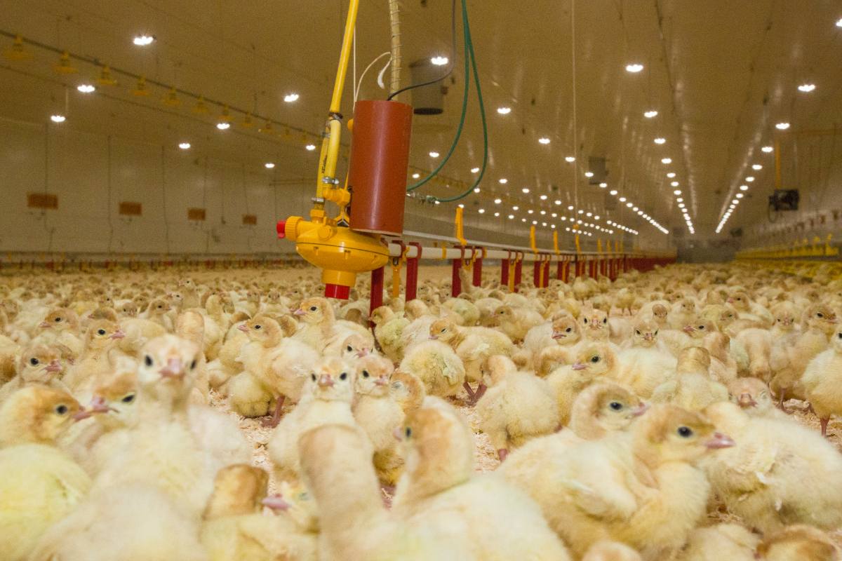
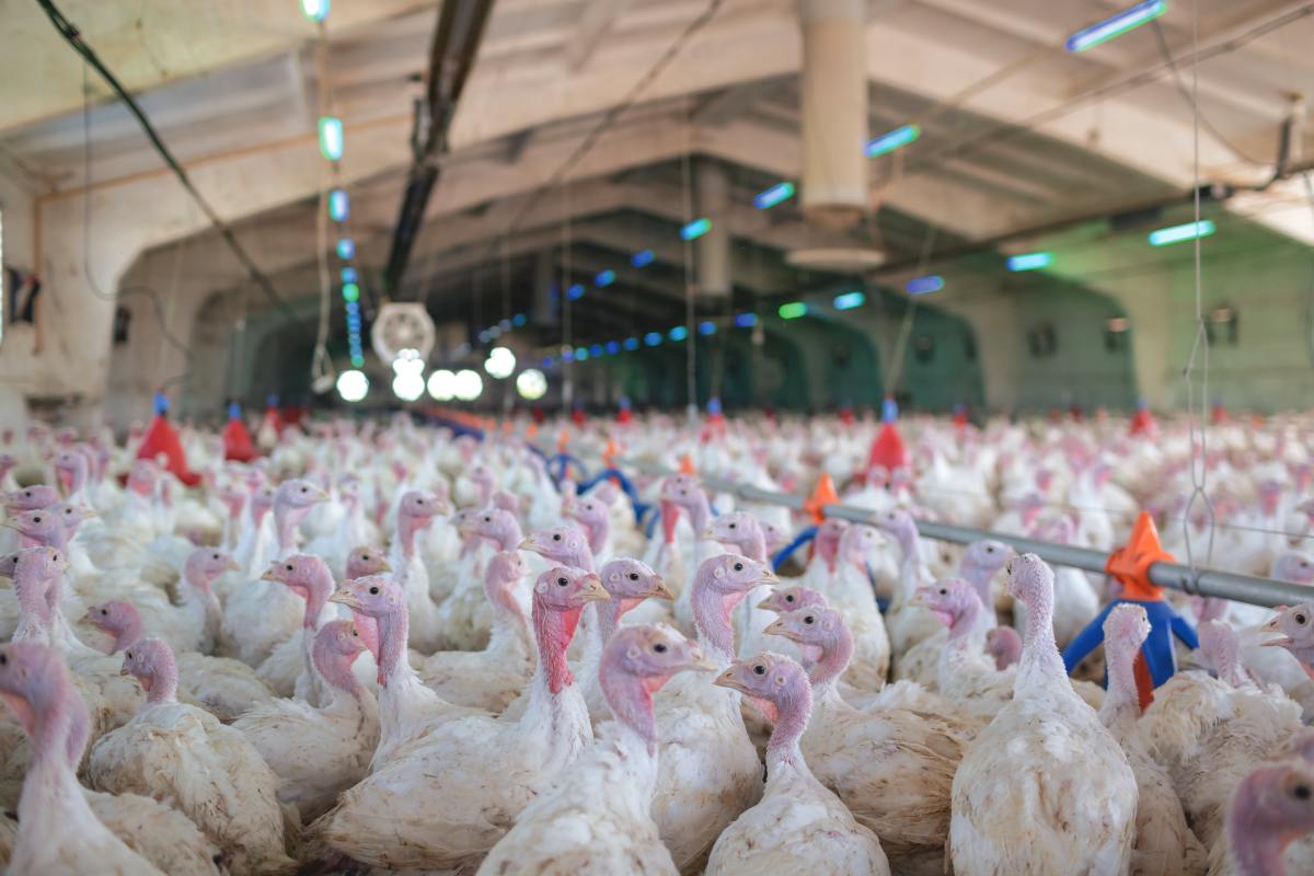
An outbreak of enteritis was reported in a commercial turkey operation. The farm consisted of four houses, each containing approximately 7,600 poults, all of which were heavy tom turkeys. The birds were raised on new litter consisting of wood shavings. At the time of sampling, the birds were 42 days of age. Clinical signs of enteric disease — including diarrhea, depressed feed intake, and poor uniformity — were observed throughout the flock, and the entire house appeared to be affected.
Intestinal scraping
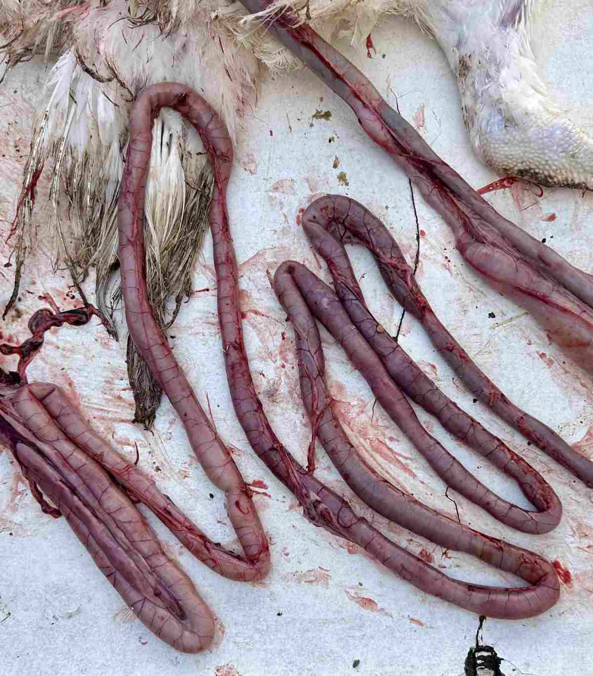
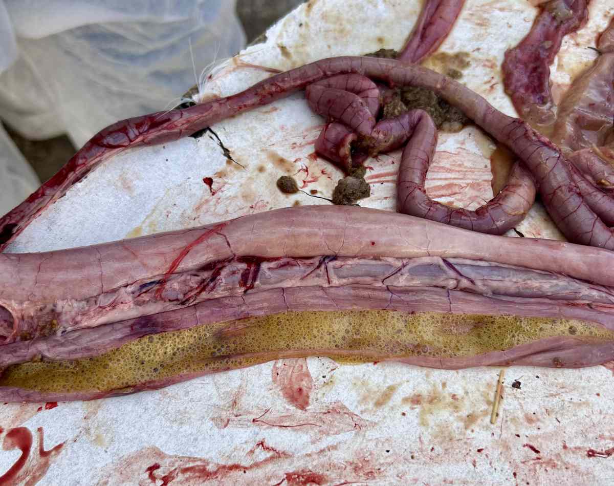
A few birds exhibiting severe clinical signs were euthanized and necropsied. Fresh mucosal scrapings were harvested from the intestines, subjected to immediate wet-mount microscopic examination and evaluated for protozoal morphology and motility.
DNA extraction and PCR
Fecal flotation was performed on pooled cecal and intestinal contents. DNA was subsequently extracted from 200 µL of post-fecal-flotation material using the QIAamp Fast DNA Stool Kit (Qiagen, Hilden, Germany), following the manufacturer’s instructions. To improve the disruption of protozoal oocysts, glass beads were incorporated into the extraction process, and 30 µL of Proteinase K (Qiagen) was added during the initial lysis/digestion step.
PCR amplification was performed using previously described primers targeting the partial 18S ribosomal RNA (rRNA) gene for trichomonads (4) and the mitochondrial cytochrome c oxidase subunit I (COI) gene for Eimeria species differentiation (5). All PCR assays were executed using GoTaq® G2 Hot Start Green Master Mix (Promega, Madison, WI) with forward and reverse primers at a final concentration of 5 µM.
Gel electrophoresis and sequencing
PCR amplicons (2 µL) were separated by electrophoresis using E-Gel™ EX Agarose Gels (2%) (Invitrogen, Carlsbad, CA) and visualized under UV illumination. A reference E-Gel™ 1 Kb Plus DNA Ladder (Invitrogen) was used to determine the size of the amplified products. Positive PCR products were purified using the QIAquick PCR Purification Kit (Qiagen) according to the manufacturer’s instructions and subsequently submitted for sequencing at Eurofins Genomics (Kentucky, USA).
 Results and discussion
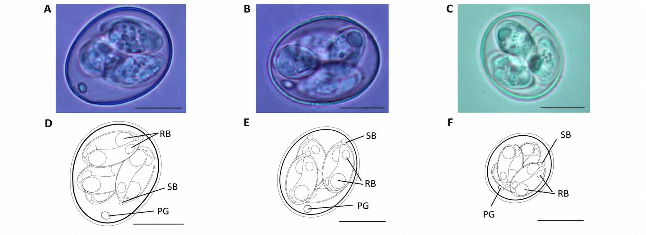
Microscopic examination of the wet-mount intestinal scrapings revealed numerous Eimeria oocysts together with highly motile, flagellated protozoa exhibiting the characteristic jerky movement indicative of Tetratrichomonas gallinarum. Molecular characterization followed by sequence analysis confirmed the flagellate as T. gallinarum. Fecal flotation similarly demonstrated an abundant shedding of Eimeria oocysts. Subsequent molecular identification and sequencing detected four Eimeria species: Eimeria adenoeides, E. meleagrimitis, E. dispersa, and E. meleagridis. Of the seven Eimeria species that infect turkeys, three are recognized as the most pathogenic: E. adenoeides, E. meleagrimitis, and E. gallopavonis (6,7,8,9). In this case, both E. adenoeides and E. meleagrimitis were identified.
The concurrent detection of T. gallinarum and multiple Eimeria species indicates a complex, mixed protozoal enteric infection. Coinfections involving coccidia and trichomonads may intensify intestinal pathology through synergistic interactions. Coccidial infection causes epithelial destruction, villus atrophy, and intestinal inflammation. This severe disruption of the mucosal barrier and subsequent impairment of nutrient absorption may alter the local microenvironment, facilitating the colonization and rapid proliferation of opportunistic enteric organisms, including trichomonads. This overgrowth can amplify the host inflammatory response and exacerbate clinical disease severity.
The pathogenic significance of T. gallinarum in turkeys remains incompletely understood and somewhat controversial. Experimental infections have generally failed to reproduce overt clinical disease or characteristic gross lesions in turkeys (10,11). Similarly, Amin et al. (12) reported that turkeys experimentally inoculated with T. gallinarum developed latent infections without significant clinical signs or pathological changes. These findings have contributed to the prevailing view that T. gallinarum possesses relatively low pathogenicity in turkeys under controlled experimental conditions.
Nevertheless, increasing evidence suggests that T. gallinarum may contribute to disease development under field conditions, particularly in the presence of concurrent infections or other predisposing factors. Crespo et al. (13) reported increased mortality and production losses in Pekin ducks coinfected with Escherichia coli and Tetratrichomonas spp., highlighting the potential for synergistic interactions between pathogens. Furthermore, T. gallinarum-associated granulomatous disease has been documented in free-range laying hens (14), while typhlitis and typhlohepatitis associated with T. gallinarum have been described in ducks (15). Extraintestinal manifestations have also been reported, including encephalitis in mockingbirds (16) and mortality events in black swans (17). Collectively, these reports indicate that although T. gallinarum may exhibit limited pathogenicity in experimentally infected turkeys. It has the potential to act as an opportunistic or contributing pathogen capable of causing significant disease in a variety of avian hosts when the intestinal ecosystem is compromised.
Among the Eimeria species identified in this clinical case, E. adenoeides and E. meleagrimitis are recognized as the most pathogenic species affecting turkeys. E. adenoeides primarily infects the ceca and is associated with severe typhlitis, hemorrhage, and characteristic cecal core formation. In severe infections, lesions may extend into the lower small intestine and cloaca (6,7,8,9). E. meleagrimitis predominantly parasitizes the duodenum and proximal jejunum, causing extensive mucosal damage, impaired nutrient absorption, and reduced growth performance (6,7,9). In contrast, E. dispersa is generally considered mildly pathogenic and produces lesions primarily in the mid-intestine, occasionally extending to the cecal necks (6,7). E. meleagridis is typically regarded as nonpathogenic, although infection of the ceca has been associated with occasional caseous cecal core formation (6,7).
Experimental evidence supports a biologically plausible interaction between coccidia and trichomonads. Norton (18) demonstrated that turkeys coinfected with E. adenoeides and T. gallinarum exhibited significantly greater cecal colonization by T. gallinarum than birds infected with the flagellate alone. These findings suggest that coccidial-induced mucosal damage may facilitate establishment, persistence, and proliferation of trichomonads within the intestinal tract. Such interactions are likely amplified under commercial production conditions where multiple infectious, nutritional, and environmental stressors are simultaneously present.
The simultaneous detection of four Eimeria species and T. gallinarum in the present case suggests that the observed enteritis was likely multifactorial and resulted from the combined effects of several protozoal pathogens rather than a single etiologic agent. The presence of multiple Eimeria species likely played a major role in initiating intestinal damage, while concurrent T. gallinarum infection may have contributed to disease progression and severity. Mixed protozoal infections have the potential to amplify intestinal injury, compromise gut integrity, reduce nutrient utilization, and increase susceptibility to secondary bacterial infections, ultimately leading to adverse effects on flock health and performance.
These findings underscore the importance of comprehensive diagnostic investigations during enteric disease outbreaks in turkeys. Relying on single-pathogen diagnostic approaches (such as visual lesion scoring alone) may underestimate the contribution of concurrent infections to disease expression and severity. Combining classical parasitological examination (wet-mounts and flotation) with molecular diagnostics (PCR and sequencing) improves the sensitivity of mixed-infection detection and facilitates an accurate etiological diagnosis. Effective control of complex enteric disease syndromes in commercial turkey production requires integrated parasite management strategies incorporating stringent biosecurity measures, routine surveillance, strategic vaccination programs where applicable, and appropriate anticoccidial interventions.
Reference
- Durairaj, V., Clark, E. B., & Vander Veen, R. (2023). Concurrent infection of Histomonas meleagridis and Pentatrichomonas hominis in a blackhead disease outbreak in turkeys. Avian Diseases, 67(1), 124–129.
- Durairaj, V., Nezworski, J., Drozd, M., Clark, S., & Vander Veen, R. (2024). Concurrent Histomonas meleagridis and hemorrhagic enteritis virus infection in a turkey flock with recurrent history of blackhead disease. Avian Diseases, 68(1), 56–64.
- Clark, S. R., & Chiaia, L. (2024). Current health issues facing the US turkey industry [PDF report]. Huvepharma, Inc. https://usaha.org/wp-content/uploads/2025/12/2024-USAHA-Proceedings-FINAL_PRINT.pdf
- Grabensteiner, E., & Hess, M. (2006). PCR for the identification and differentiation of Histomonas meleagridis, Tetratrichomonas gallinarum and Blastocystis spp. Veterinary Parasitology, 142(3–4), 223–230.
- Hafeez, M. A., Shivaramaiah, S., Dorsey, K. M., Ogedengbe, M. E., El-Sherry, S., Whale, J., Cobean, J., & Barta, J. R. (2015). Simultaneous identification and DNA barcoding of six Eimeria species infecting turkeys using PCR primers targeting the mitochondrial cytochrome c oxidase subunit I (mtCOI) locus. Parasitology Research, 114, 1761–1768.
- Lund, E. E., & Farr, M. M. (1965). Coccidiosis of the turkey. In H. E. Biester & L. H. Schwarte (Eds.), Diseases of poultry (5th ed., pp. 1088–1093). Iowa State University Press.
- Cervantes, H. M., McDougald, L. R., & Jenkins, M. C. (2020). Coccidiosis. In D. Swayne, M. Boulianne, C. Logue, L. McDougald, V. Nair, D. Suarez, S. deWit, T. Grimes, D. Johnson, M. Kromm, et al. (Eds.), Diseases of poultry (14th ed., pp. 1193–1216). Wiley-Blackwell.
- Durairaj, V., & Vander Veen, R. (2025, March). Cecal coccidiosis in turkeys. Zootecnica, 40–42.
- Durairaj, V., & Vander Veen, R. (2025, April). Pathological manifestations of highly pathogenic and predominant Eimeria species in turkeys. Zootecnica, 38–42.
- Delappe, I. P. (1957). Effect of inoculating the chicken and the turkey with a strain of Trichomonas gallinarum. Experimental Parasitology, 6(4), 412–417.
- Pecka, Z. (1991). Pathogenicity of Tetratrichomonas gallinarum. Veterinární Medicína (Praha), 36(3), 183–188.
- Amin, A., Liebhart, D., Weissenböck, H., & Hess, M. (2011). Experimental infection of turkeys and chickens with a clonal strain of Tetratrichomonas gallinarum induces a latent infection in the absence of clinical signs and lesions. Journal of Comparative Pathology, 144(1), 55–62. https://doi.org/10.1016/j.jcpa.2010.06.002
- Crespo, R., Walker, R. L., Nordhausen, R., Sawyer, S. J., & Manalac, R. B. (2001). Salpingitis in Pekin ducks associated with concurrent infection with Tetratrichomonas sp. and Escherichia coli. Journal of Veterinary Diagnostic Investigation, 13(3), 240–245.
- Landman, W. J. M., Gantois, N., van Eck, J. H. H., van der Heijden, H. M. J. F., & Viscogliosi, E. (2019). Tetratrichomonas gallinarum granuloma disease in a flock of free-range layers. Veterinary Quarterly, 39(1), 153–160. https://doi.org/10.1080/01652176.2019.1682714
- Richter, B., Schulze, C., Kämmerling, J., Mostegl, M., & Weissenböck, H. (2010). First report of typhlitis/typhlohepatitis caused by Tetratrichomonas gallinarum in three duck species. Avian Pathology, 39(6), 499–503.
- Patton, C. S., & Patton, S. (1996). Tetratrichomonas gallinarum encephalitis in a mockingbird (Mimus polyglottos). Journal of Veterinary Diagnostic Investigation, 8(1), 133–137.
- Feng, S., Chang, H., Wang, Y., Luo, F., Wu, Q., Han, S., & He, H. (2021). Lethal infection caused by Tetratrichomonas gallinarum in black swans (Cygnus atratus). BMC Veterinary Research, 17(1), 191. https://doi.org/10.1186/s12917-021-02894-x
- Norton, R. A. (1997). Pathogenicity of a strain of Trichomonas gallinarum in turkeys and its possible interaction with cecal coccidia. Avian Diseases, 41(3), 670–675.
Apa Reaksi Anda?
 Suka
0
 Kurang Suka
0
 Setuju
0
 Tidak Setuju
0
 Bagus
0
 Berguna
0
 Hebat
0