




















































Tissue Engineering Takes Shape

Tissue engineering is on the move from an ambitious idea toward everyday clinical reality. At the interface of biology, materials science, and engineering, the field seeks to repair or replace damaged human tissues and organs. Some approaches embrace personalized medicine, creating tissues and treatments from a patient’s own cells and materials. Others are developing off-the-shelf engineered products for broad availability. We spoke to experts in the field about the latest developments pushing both strategies forward. Technologies range from heart patches and spinal cord repair to bone, cartilage and vascular grafts, and beyond.
Living cardiac band-aid

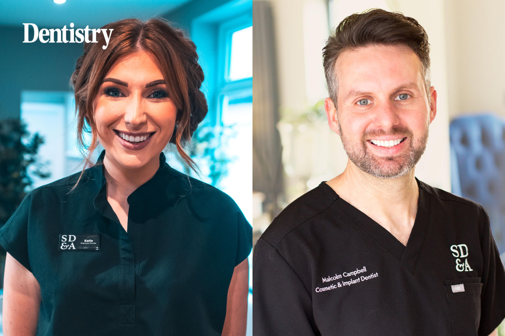
CEO, Avery Therapeutics
“The concept of making functional tissue to replace damaged tissues and organs has captured the imagination of many for a long time,” says Jordan Lancaster, PhD, CEO of Avery Therapeutics. Case in point: heart failure. Lancaster elaborates, “Today, we manage cardiac diseases like heart failure by administering drugs that help reduce symptoms of the disease. And while the current armament of drugs for heart failure does slow progression of disease, the prognosis remains grim, and the disease continues to advance. There are no therapies today that get to the root cause of heart failure, which is loss of functioning cardiomyocytes (heart muscle cells) and fibrosis. We want to change this. We want to develop novel therapies that address the root cause of disease, and reverse disease in ways that are currently unobtainable.”
According to Lancaster, the company is pursuing an immunomodulatory approach coupled with tissue engineering to essentially create a “living band-aid, of sorts.” Their preclinical collaborative studies employed an allogeneic cardiac patch composed of a proprietary mix of human induced pluripotent stem cell (iPSC)-derived cardiomyocytes and fibroblasts embedded in a bioresorbable matrix. Lancaster explains, “This biologic product is placed on damaged tissue and creates a localized immunomodulatory response that drives disease repair. Our immunomodulatory platform is built utilizing cellular material, a proprietary combination of cells produced via a multi-day manufacturing process. The biologic engages the immune system and recruits immune precursor cells, which drive repair.”
Developing the approach also required overcoming key hurdles such as manufacturing at scale, handleability, and shelf stability (cryopreservation). Additionally, Lancaster says the company has learned a lot about the immune system and its ability to serve as a conserved therapeutic target for diseases of inflammation and fibrosis. “We are exploring and have established deep mechanistic insight into how to redirect the immune system into pro-reparative pathways, which we have validated in highly translatable preclinical models of heart failure. We are keen to advance these insights into novel therapies.”
As a late-stage preclinical company, Avery will initially focus on heart failure of ischemic etiology with plans to advance the technology toward clinical testing.
Bioinks for 3D bioprinting

CEO, CollPlant Biotechnologies
While tissue engineering has made remarkable progress, fundamental barriers remain, according to Yehiel Tal, CEO of CollPlant Biotechnologies, including “access to safe and reproducible biomaterials, recreation of the complex biological and mechanical environment of human tissue, vascularization, and scalable manufacturing.” Bioinks, specialized cell-laden gel-based biomaterials, are a critical aspect. He elaborates, “Bioinks must support cell viability while providing the printability, structural stability, degradation profile, and mechanical properties required for functional tissue formation.”
The company is addressing these challenges through its proprietary plant-derived recombinant human type 1 collagen (rhCollagen) platform. Tal reports, “Because our collagen is bioidentical to human collagen and produced without animal or human tissue, it offers a consistent, scalable, animal-free building block for regenerative medicine. Our bioink portfolio combines rhCollagen with complementary biomaterials and photoactive components to tailor mechanical strength, degradation kinetics, and printing performance for different tissues and printing technologies.”

One example from this portfolio is BioFlex , a ready-to-print kit for high-resolution digital light processing (DLP) bioprinting. DLP bioprinting projects a digital image of each layer of a desired 3D structure onto light-sensitive bioink, selectively solidifying the material to build the structure layer by layer. Tal explains, “BioFlex combines Collink.3D 50 with a biodegradable polymer and proprietary photoactive agents, simplifying formulation while retaining the flexibility to create complex, biologically relevant structures. It is intended for advanced tissue models, drug discovery, engineered tissues, and ultimately transplantable organs.”
The company is also applying tissue engineering to medical aesthetics. Tal says, “We have advanced commercial-size, 3D-bioprinted regenerative breast implants through large-animal preclinical studies. These implants are designed to gradually degrade while supporting vascularization and regeneration of the patient’s own tissue.”
Tal believes progress in the field will depend on integrating biomaterials, cells, vascular networks, bioreactors, automation, and computational design into scalable manufacturing platforms. “We see major opportunities in human tissue models, regenerative soft-tissue products, and organ manufacturing, enabled by biomaterials that are both biologically relevant and industrially reproducible.”
Personalized spinal cord repair
More than 18,000 people sustain traumatic spinal cord injuries in the U.S. annually, with approximately 310,000 patients living with the condition. The injured spinal cord has little regenerative capacity and develops a cellular and extracellular environment that inhibits neural regrowth.

CEO, Matricelf
Gil Hakim, CEO of Matricelf, reports, “Chronic spinal cord injury is characterized by tissue cavities and scarring that interrupt neural pathways. Regeneration requires addressing inflammation, restoring extracellular-matrix cues, and enabling transplanted cells to survive, organize, extend axons, form functional synapses, and integrate with spinal cord circuitry.”
Matricelf is developing NewRal, a 3D neural-tissue transplant composed of patient blood-derived iPSCs as well as an extracellular-matrix hydrogel derived from the patient’s omental tissue. Before transplantation, the cells differentiate and assemble into 3D neural tissue spheroid constructs containing electrically active neural cells, designed to replace damaged neural tissue and support new neural connections.
“The dual-autologous design may reduce immune mismatch and the need for immunosuppression,” says Hakim. “Compared with transplants using donor cells or synthetic, animal-derived, or standardized scaffolds, NewRal combines patient-specific cells, biological matrix, and preassembled neural tissue.”
Matricelf is addressing manufacturing complexity and patient variability through standardized Good Manufacturing Practices (GMP) processes and optimization via closed-system automation. Hakim indicates that pilot efficacy studies in chronic spinal cord injury animal models have shown improvement in functional recovery and tissue regeneration in 80% of treated animals. The company completed Good Laboratory Practice (GLP) safety studies, advanced GMP manufacturing and clinical site preparations, initiated patient identification activities, and received FDA feedback on elements of the proposed first-in-human clinical program planned for 2027.

Living bone and cartilage grafts

CEO, EpiBone
Tissue engineering is also making strides in the repair of bone and cartilage. According to Nina Tandon, PhD, CEO of EpiBone, “The gold standard treatments for skeletal defects are unfortunately riddled with compromises to meet the standard of ‘replacing like with like.’ Craniofacial bone loss after trauma or tumor surgery, and damaged joint cartilage, are the big ones. What happens today? A surgeon can harvest bone from your hip or leg, so now you have two wounds and real donor-site pain. They can use cadaver tissue, which the body often resorbs. Or they can bolt in metal or plastic that never becomes bone and, sooner or later, needs revising.”
EpiBone aims to shift from replacing tissue to regrowing it using stem cells to create personalized bone and cartilage products. Tandon explains, “Depending on the case, we can shape a graft to a patient’s exact anatomy before it reaches the operating room, or supply it off the shelf, ready when the surgeon needs it. Either way, it is living tissue on a natural bone matrix, so it is designed to integrate and remodel the way native tissue does rather than be tolerated as a foreign object.”
The process begins with adult stem cells seeded onto a natural bone matrix and matured in a bioreactor. Tandon elaborates, “I describe the bioreactor as diet and exercise: it feeds the tissue and puts it through the mechanical loading it will see in the body (going jogging a couple miles a day, roughly). Out comes living, 3D bone or cartilage. Honestly, the biology is the fun part. The moat is manufacturing. Making living tissue reproducibly, at the right size and shape and at clinical grade, is the hard problem, and it is the one we have spent more than a decade on.”
The company has advanced its bone and cartilage programs into human trials. Tandon reports, “In our first-in-human bone trial, we treated six patients with zero product-related adverse events. Those six jaws are the thing I am proudest of.” EpiBone has also tested an injectable cartilage therapy for osteoarthritis in dogs and horses, reporting improvements over standard-of-care treatment.
Looking ahead, Tandon sees the next decade focusing on scale and broader availability of custom and off-the-shelf living grafts.
Off-the-shelf vascular grafts
When a patient suffers a severe injury or chronic vascular disease, surgeons may need to replace or repair an artery. Current options include using a patient’s own vein, synthetic grafts, or an animal-derived allograft.

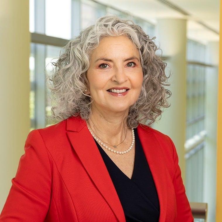
President and CEO, Humacyte
“Harvesting a vein can be especially dangerous in urgent cases, taking more time in the OR, adding increased risk for infection, and resulting in a second surgical wound with the potential for post-operative pain,” says Laura Niklason, MD, PhD, president and CEO of Humacyte. She also notes some synthetic grafts are prone to infection while animal-derived allografts carry other issues. What is needed is a readily available conduit that behaves like living tissue but resists infection, maintains patency, and integrates seamlessly with the body.
Humacyte has developed acellular tissue-engineered vessels (ATEVs) as off-the-shelf, universally implantable vascular grafts. Niklason explains, “We begin with human vascular smooth muscle cells seeded onto a biodegradable scaffold, which gradually dissolves as the tissue forms.” Pulsatile, artery-like conditioning then prompts the cells to build a strong matrix of human collagen and other extracellular matrix proteins. After the vessel matures, the cells used to grow it are removed while the extracellular matrix and structural proteins remain. The resulting bioengineered vessel can be stored until needed. After implantation, the patient’s own cells gradually repopulate and remodel the vessel, transforming it into living tissue.
The company’s lead product, Symvess®, was approved by the FDA in December 2024 as a vascular conduit for adults with extremity arterial injury when urgent revascularization is needed to avoid imminent limb loss and an autologous vein graft is not feasible. In September 2026, the FDA approved extending its shelf life from 18–36 months. Humacyte is also planning submission of a supplemental Biologics License Application in the second half of 2026 for hemodialysis applications.
Niklason believes tissue engineering is at an important inflection point. “What’s changing now is the shift toward manufactured, off-the-shelf regenerative products that have the potential to reach a large scale of patients. The opportunity ahead is to make engineered human tissue a routine part of surgical and medical care for the benefit of the patients who need it.”
The post Tissue Engineering Takes Shape appeared first on GEN - Genetic Engineering and Biotechnology News.
Apa Reaksi Anda?
 Suka
0
 Kurang Suka
0
 Setuju
0
 Tidak Setuju
0
 Bagus
0
 Berguna
0
 Hebat
0